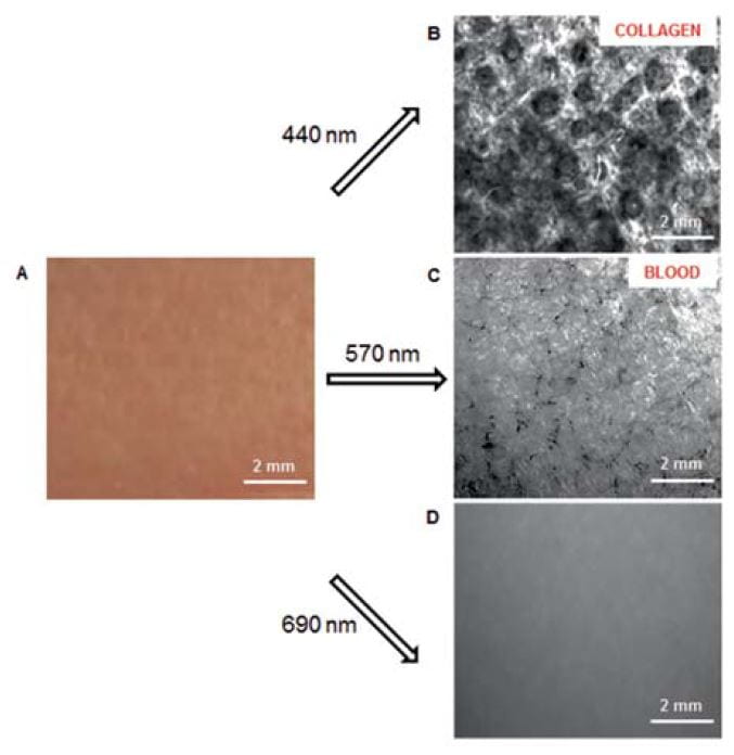
Optical techniques can be used to monitor changes in morphology, biochemical and biophysical properties of skin that may indicate aging, pathological processes, and even development of skin cancer. Real-time, non-invasive monitoring and quantitative assessment of skin structures in vivo is of paramount importance because skin cancer is the most common malignancy in the United States. In addition, we have introduced and validated multimodal optical imaging methods for evaluation of brain structure and function.
Selected Publications:
Feng X, Doherty S, Yaroslavsky I, Altshuler G, and Yaroslavsky AN, “Polarization enhanced wide-field imaging for evaluating dermal changes caused by non-ablative fractional laser treatment,” Lasers Surg Med. 48(2), 150-156 (2016).
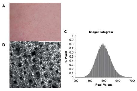
Feng X, Patel R, and Yaroslavsky AN, “Wavelength optimized cross-polarized wide-field imaging for noninvasive and rapid evaluation of dermal structures,” J Biophotonics. (2015).
Park J, Mroz P, Hamblin MR, and Yaroslavsky AN, “Dye-enhanced multimodal confocal microscopy for noninvasive detection of skin cancers in mouse models,” J.Biomed. Opt. 15(2), 026023 (2010).