The goal of this research is the development of devices and methods for the detection of cancer at the cellular level. Our in-house built multi-modal confocal microscopy systems provide high resolution, high contrast images that permit discrimination of benign and malignant cells based on morphology and quantitative measurements.
Selected Publications:
Feng X, Muzikansky A, Ross M., Hamblin M., Jermain P., and Yaroslavsky AN, “Multimodal quantitative imaging of brain cancer in cultured cells,” Biomed. Opt. Express 10(8), 4237 (2019).
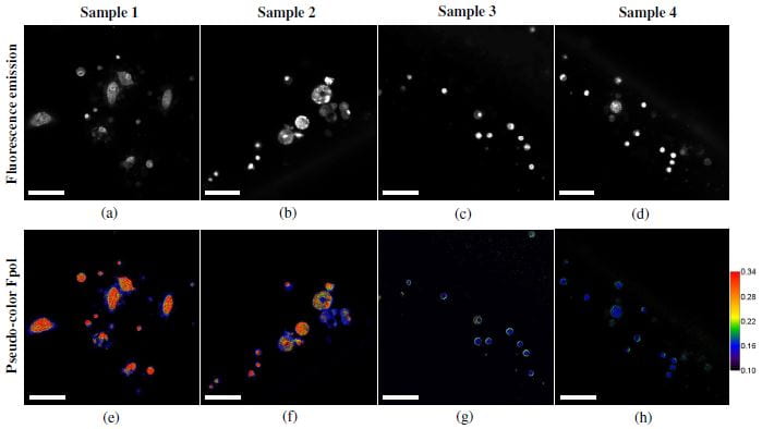
Malik S, Jermain P, Feng X, and Yaroslavsky AN, “Multimodal optical imaging of renal cells,” Opt Eng. 58(8), (2019).
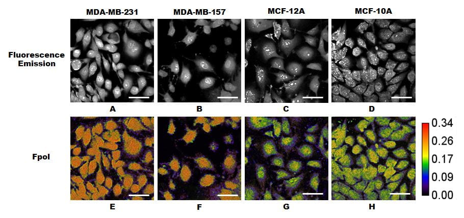
Yaroslavsky AN, Feng X, Muzikansky A, and Hamblin M, “Fluorescence polarization of methylene blue as a quantitative marker of breast cancer at the cellular level,” Scientific Reports. 9(1), (2019).
Wirth D, Smith TW, Moser R, and Yaroslavsky AN, “Demeclocycline as a contrast agent for detecting brain neoplasms using confocal microscopy,” Phys Med Biol. 60(7), 3003-3011 (2015).
Wirth D, Snuderl M, Curry W, and Yaroslavsky A, “Comparative evaluation of methylene blue and demeclocycline for enhancing optical contrast of gliomas in optical images,” J. Biomed. Opt. 19(9), (2014).
Snuderl M, Wirth D, Sheth S, Bourne S, Kwon C, Curry WT, Frosch MP, and Yaroslavsky AN, “Dye-enhanced multimodal confocal imaging as a novel approach to intraoperative diagnosis of brain tumors,” Brain Pathol. 23(1), 73-81 (2013).
Kamionek M, Yaroslavsky AN, Patel R, Kandil D, Wirth D, Quinlan R, and Khan A, “Feasibility of dye-enhanced fluorescence confocal microscopy in differentiation of benign and malignant breast tissue,” Am J Clin Pathol. 138, A367 (2012).
Wirth DJ, Snuderl M, Sheth M, Kwon C, Frosch MP, Curry W, and Yaroslavsky AN, “Identifying brain neoplasms using dye-enhanced multimodal confocal imaging,” J.Biomed. Opt. 17(20), (2012).